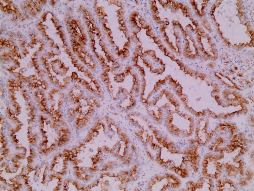
Recognizes a ~200kDa glycoprotein (gp200) which is identified as a renal cell carcinoma marker . Its epitope resides in the carbohydrate domain of gp200 . It shows no significant cross-reactivity with other carbohydrate determinants, such as the Lewis blood group antigens, epithelial membrane antigen, HMFG, and AB blood group antigens . In normal kidney, gp200 is localized along the brush border of the pars convoluta and pars recta segments of the proximal tubule, as well as focally along the luminal surface of Bowman's capsule adjoining the outgoing proximal tubule . Of other normal tissues examined, the gp200 is also localized along the luminal surfaces of breast lobules and ducts, the luminal surface of the epididymal tubular epithelium, within the cytoplasm of parathyroid parenchymal cells, and focally within the colloid of thyroid follicles. Thirty-one other normal tissues failed to express similar or cross-reacting antigens . Reportedly, gp200 is expressed by 93% of primary and 84% of metastatic renal cell carcinomas . This antibody may be a useful reagent in the investigations of carcinomas of proximal nephrogenic differentiation especially those showing tubular differentiation. Absence of reactivity with alpha fetoprotein (AFP) and alpha-1-antitrypsin antibodies and exclusion of morphological or clinical criteria which might otherwise indicate metastatic breast or embryonal carcinoma would be further confirmator.